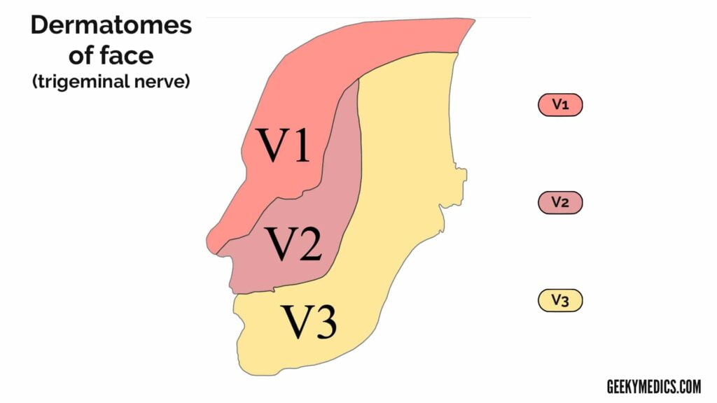
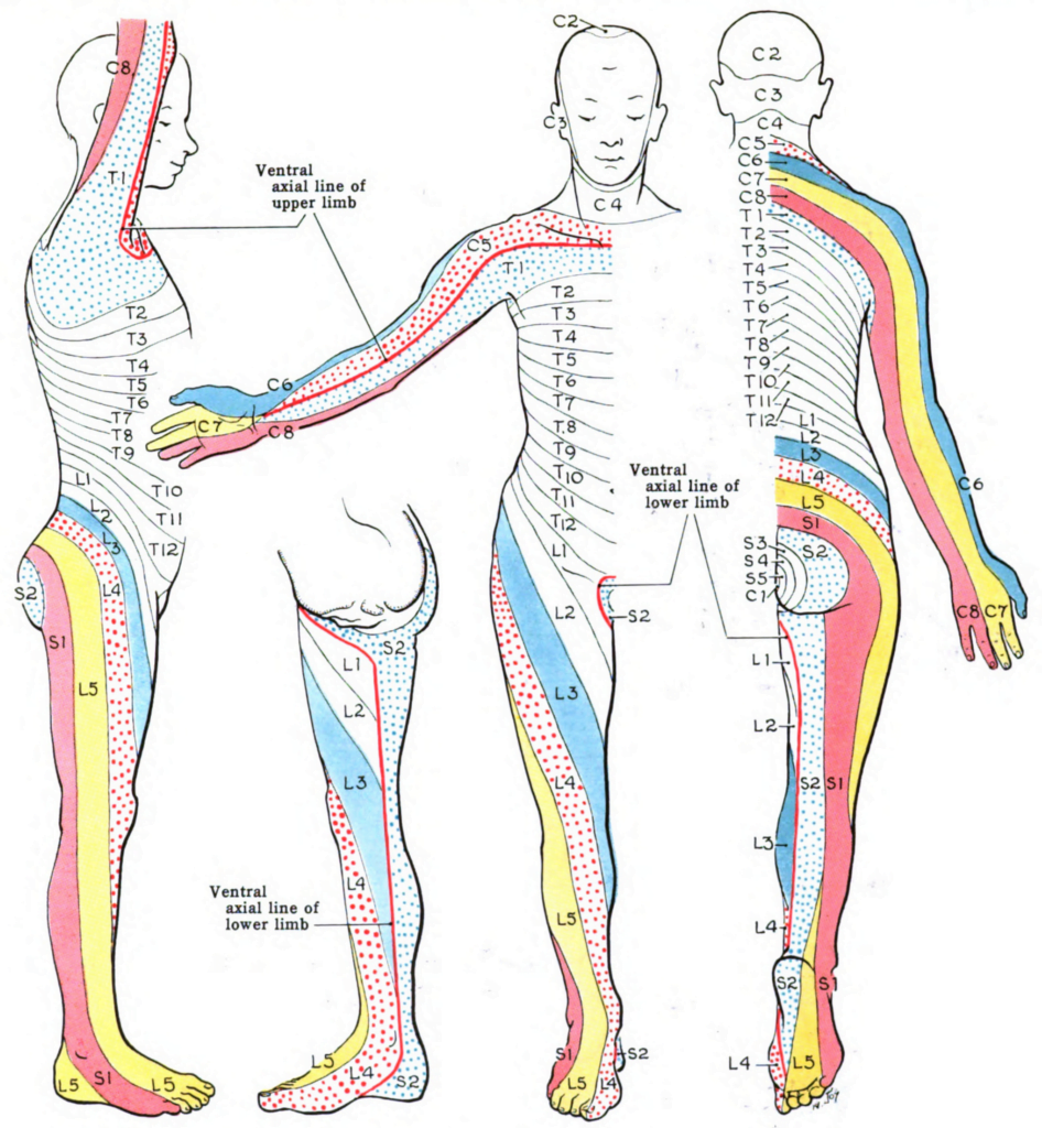
Dermatomes Of Trigeminal Nerve – A dermatome is the location of the skin of the human anatomy that is mainly supplied by branches of a single back sensory nerve root. These back sensory nerves get in the nerve root at the spinal cord, and their branches reach to the periphery of the body. The sensory nerves in the periphery of the body are a type of nerve that transmits signals from experiences (for instance, pain symptoms, touch, temperature) to the spine from particular locations of our anatomy.
Why Are Dermatomes Most important?
To comprehend dermatomes, it is essential to understand the anatomy of the spinal column. The spine is divided into 31 segments, each with a set (right and left) of posterior and anterior nerve roots. The kinds of nerves in the anterior and posterior roots are various. Anterior nerve roots are responsible for motor signals to the body, and posterior nerve roots receive sensory signals like discomfort or other sensory symptoms. The anterior and posterior nerve roots combine on each side to form the back nerves as they leave the vertebral canal (the bones of the spinal column, or backbone).
Dermatomes And Myotomes Sensation Anatomy Geeky Medics
Dermatomes And Myotomes Sensation Anatomy Geeky Medics
Dermatome charts
Dermatome maps illustrate the sensory distribution of each dermatome throughout the body. Clinicians can evaluate cutaneous feeling with a dermatome map as a method to localise sores within central nervous tissue, injury to particular back nerves, and to identify the degree of the injury. Several dermatome maps have been developed for many years but are often clashing. The most typically used dermatome maps in significant books are the Keegan and Garrett map (1948) which leans towards a developmental interpretation of this idea, and the Foerster map (1933) which associates better with medical practice. This post will examine the dermatomes using both maps, determining and comparing the major distinctions between them.
It’s very important to tension that the existing Dermatomes Of Trigeminal Nerve are at best an estimation of the segmental innervation of the skin considering that the many locations of skin are generally innervated by a minimum of two spinal nerves. For example, if a patient is experiencing numbness in only one location, it is unlikely that numbness would take place if only one posterior root is impacted because of the overlapping division of dermatomes. A minimum of two neighboring posterior roots would need to be impacted for numbness to occur.
Dermatome Anatomy Wikipedia
Dermatome anatomy Wikipedia
The Dermatomes Of Trigeminal Nerve typically play an essential function in figuring out where the problem is originating from, giving doctors a hint regarding where to look for indications of infection, swelling, or injury. Typical diseases that may be partially identified through the dermatome chart include:
- Spinal injury (from a fall, etc.)
- Compression of the spinal cord
- Pressure from a tumor
- A hematoma (pooling blood)
- Slipped or bulging discs
A series of other analysis devices and signs are very important for identifying injuries and diseases of the spine, including paralysis, bladder dysfunction, and gait disruption, along with analysis procedures such as imaging (MRI, CT, X-rays checking for bone damage) and blood tests (to look for infection).
Dermatomes play a significant role in our understanding of the human body and can assist clients much better comprehend how issue to their back can be identified through different signs of discomfort and other unusual or out-of-place experiences.Dermatomes Of Trigeminal Nerve
When the spine is damaged, treatments frequently include medication and intervention to decrease and combat swelling and workout, swelling and rest to minimize discomfort and reinforce the surrounding muscles, and in specific cases, surgical treatment to remove bone spurs or pieces, or decompress a nerve root/the spine.Dermatomes Of Trigeminal Nerve

